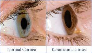
Keratoconus occurs when your cornea — the clear, dome-shaped front surface of your eye — thins and gradually bulges outward into a cone shape.
The cornea is the clear, outer layer at the front of your eye.
The middle layer is the thickest part of the cornea, mostly made up of water and a protein called collagen. Collagen makes the cornea strong and flexible, and helps keep its regular, round shape.
This healthy cornea focuses light so you can see clearly. With keratoconus, the cornea thins and bulges into an irregular cone shape, resulting in vision loss.
hopkinsmedicine.org
It can affect one or both eyes (usually one more than the other) and normally progesesses slowly.
There is no way to predict how quickly the disease will progress, or if it will progress at all.
In the early stages of keratoconus, you might be able to correct vision problems with glasses or soft contact lenses.
Later, you may have to be fitted with rigid, gas permeable contact lenses or other types of lenses, such as scleral lenses.
If your condition progresses to an advanced stage treatments such as corneal collagen cross-linking and/or and/or corneal ring and /or you may need a cornea transplant.
Mayoclinic
Diagnosis
Corneal topography. This is the most accurate way to diagnose early keratoconus and follow its progression. A computerized image is taken that creates a map of the curve of the cornea.
Slit-lamp exam. This examination of the cornea can help detect abnormalities in the outer and middle layers of the cornea.
Pachymetry. This test is used to measure the thinnest areas of the cornea.
hopkinsmedicine.org
Cause
Unknown
Maybe caused by some imbalance between production and destruction of the corneal tissue by the corneal cells at birth.
Risk factors:
Having a family history of keratoconus
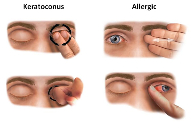
Rubbing your eyes vigorously
Constant inflammation from allergies or irritants can contribute to the destruction of corneal tissue that may result in developing keratoconus.
Having certain conditions:
Retinitis pigmentosa – a genetic condition where cells in the retina break down slowly over time.
One should have routine eye tests at least every two years but if one has concerns one should book an eye test at any point
Persons over 40 years should have eye test at least every 2 years and you must tell drivers authority if it affects your driving
Exercise your eyes
Focus change
This exercise works by challenging your focus. It should be done from a seated position.
Hold your pointer finger a few inches away from your eye.
Focus on your finger.
Slowly move your finger away from your face, holding your focus.
Look away for a moment, into the distance.
Focus on your outstretched finger and slowly bring it back toward your eye.
Look away and focus on something in the distance.
Repeat three times.
Near and far focus
This is another focus exercise.
As with the previous one, it should be done from a seated position.
Hold your thumb about 10 inches from your face and focus on it for 15 seconds.
Find an object roughly 10 to 20 feet away, and focus on it for 15 seconds.
Return your focus to your thumb.
Repeat five times.
Figure eight
This exercise should be done from a seated position as well.
Pick a point on the floor about 10 feet in front of you and focus on it.
Trace an imaginary figure eight with your eyes.
Keep tracing for 30 seconds, then switch directions.
20-20-20 rule
Eye strain is a real problem for a lot of people. Human eyes are not supposed to be glued to a single object for extended periods of time. If you work at a computer all day, the 20-20-20 rule may help prevent digital eye strain.
To implement this rule, every 20 minutes, look at something 20 feet away for 20 seconds.
Vision therapy
Vision therapy may include eye exercises, but only as part of a more specialised treatment program done under the supervision of an eye doctor, optometrist, or ophthalmologist.
The goal of vision therapy can be to strengthen the eye muscles.
It also can help to retrain poor visual behaviour, or help with eye tracking issues.
Healthline : Medically reviewed by Ann Marie Griff, O.D. — Written by Corinne O’Keefe Osborn — Updated on September 29, 2018
Please talk to your healthcare professional (i.e. Medical Doctor/Pharmacist/Optician) for further advice
Detailed Information
Please copy and paste any key words from the title: Keratoconus in the following respective 'Medtick References and/or Sources' to find out more about the disease (this also may include diagnosis tests and generic medical treatments).
The Pharmaceutical Journal covers analysis, features, opinion, learning and careers articles, providing insight and knowledge about drugs, pharmacy practice, medicines use and healthcare policy in the context of the pharmacy profession and pharmaceutical science.